Fig. 1.
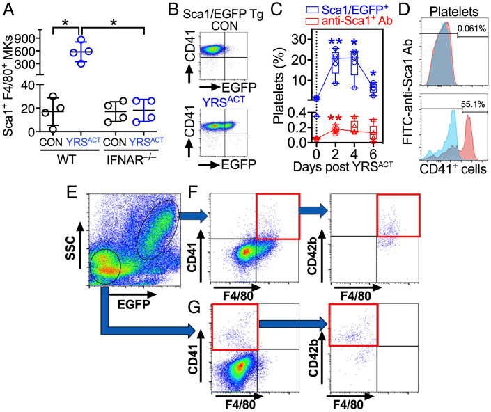
Characterization of Sca1/EGFP+ MKs and platelets. (A) BM cells from WT or IFNAR−/− mice were cultured with 200 nM YRSACT or vehicle control (CON). After 3 d, CD41+ cells costained by anti-Sca1 and anti-F4/80 antibodies were enumerated by flow cytometry (Sca1+F4/80+ MKs). Data are shown as scatter plots with mean ± SD (n = 4). (B) Flow cytometry of blood platelets from Sca1/EGFP Tg mice treated with 30-mg/kg intravenous YRSACT or CON. (C) Percentage of blood platelets from the above mice showing green fluorescence (Sca1/EGFP+; blue) or anti-Sca1 binding (red) measured at indicated times after treatment. Data (n = 5), shown as 25th–75th percentile boxes with min-to-max whiskers, were analyzed by (A) Brown-Forsythe/Welch one-way ANOVA with Dunnett’s T3 posttest for paired comparisons; or (C) repeated measures (RM) two-way ANOVA without assuming sphericity (Geisser–Greenhouse correction) with Dunnett’s posttest for before–after comparisons. *P <0.05, **P <0.01. (D) Anti-Sca1 antibody binding is negligible in blood platelets (Top), but present in >50% CD41+ BM cells (Bottom). (E) BM cells from Sca1/EGFP Tg mice gated as Sca1/EGFP − or + by flow cytometry (circled, Bottom Left or Upper Right, respectively). (F) Sca1/EGFP+ cells included CD41+, F4/80+ and CD41+F4/80+ (double-positive) cells (Left); ~half of the latter was also CD42b+ (Right). (G) Sca1/EGFP− cells included CD41+ and F4/80+ cells, but double-positive cells were negligible (Left); over half of the CD41+ cells was also CD42b+ (Right).

