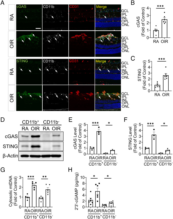
Fig. 2.
cGAS-STING signaling was overactivated in retinal myeloid cells in the OIR model. A: Representative images of immunostaining for cGAS, STING, CD31, and CD11b in retinal cryosections of room air (RA) controls and OIR mice at P17. The white arrows indicated the cGAS and STING staining co-localized with CD11b staining. GCL: ganglion cell layer, IPL: inner plexiform layer, INL: inner nuclear layer, ONL: outer nuclear layer, and OPL: outer plexiform layer. (Scale bar: 50 µm.) B and C: cGAS and STING immunostaining intensities in (A) were quantified by ImageJ software (n = 6). D: Representative western blots of cGAS and STING in myeloid cells (CD11b+) and non-myeloid cells (CD11b−) isolated from RA and OIR retinas at P17 using MACS separation. E and F: Protein levels of cGAS (E) and STING (F) in (D) were quantified by densitometry and normalized by β-actin levels (n = 3). G: Cytosolic mtDNA levels in isolated CD11b+cells were quantified by qPCR analysis using primers for a mitochondrial gene mt-Co1 (n = 5). H: Levels of 2′3′-cGAMP were measured in the CD11b+ and CD11b− cells of RA and OIR retinas at P17 using 2′3′-cGAMP ELISA kit (n = 6). Data were presented as mean ± SEM. *P < 0.05, **P < 0.01, ***P < 0.001.

