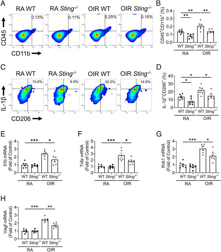
Fig. 4.
Knockout of Sting attenuated the overactivation of retinal myeloid cells in the OIR model. A: Representative flow cytometric plots of retinal myeloid cells (CD45+CD11b+) in the retinas of WT and Sting−/− mice in RA control and OIR groups at P17. B: Flow cytometric quantification of retinal myeloid percentage in the retinal cells in (A) (n = 6). C: Representative flow cytometric plots of IL-1β+CD206+ in CD45+CD11b+ cells in the retinas of RA controls and OIR mice at P17. D: Flow cytometric quantification of IL-1β+CD206+ fractions in retinal CD45+CD11b+ cells in (C) (n = 6). E–H: qRT-PCR analysis of Il1b, Tnfα, Ifnb1, and Vegf mRNA levels in MACS-isolated CD11b+ retinal cells at P17 (n = 6). Data were presented as mean ± SEM. *P < 0.05, **P < 0.01, ***P < 0.001.

