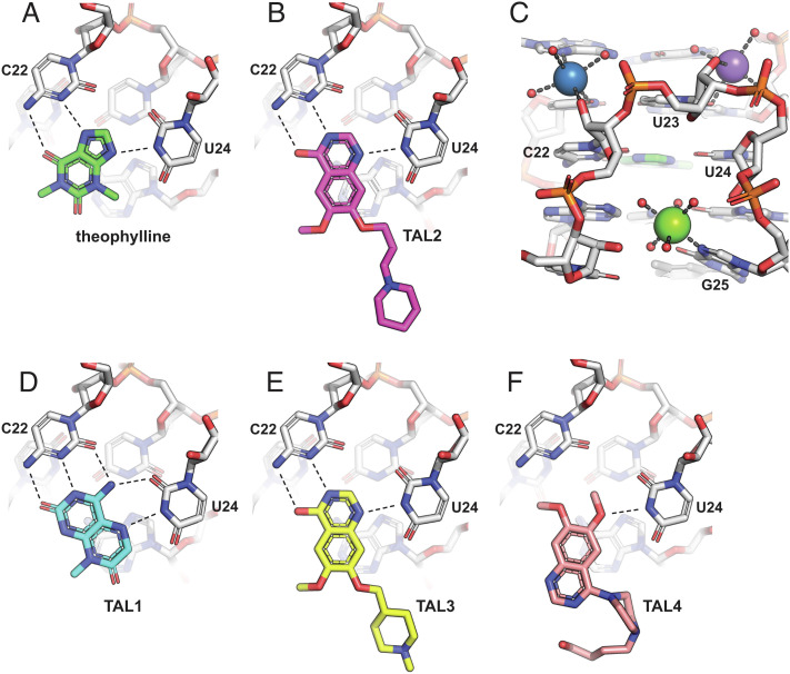
Fig. 4.
X-ray crystal structures of the theophylline aptamer with various bound ligands. (A) Theophylline forms a base triple with C22 (two hydrogen bonds) and U24 (one hydrogen bond), which stacks between two other base triples (A7–C8–G26 and U6–U23–A28). (B) TAL2 binds similarly to theophylline. (C) The binding pocket is stabilized by a Mg2+ ion (green sphere) that coordinates to N7 of G25, a Na+ ion (blue sphere) that coordinates to the 2′-hydroxyl of C22, and a second Na+ ion (purple sphere) that coordinates with U23 and forms water-mediated interactions with the sugar-phosphate of U24. Ion-coordinated water molecules are shown as red spheres. (D) TAL1 binds in the same pocket but with a third hydrogen bond to C22 and a second hydrogen bond to U24. (E) TAL3 binds similarly to TAL2. (F) TAL4 binds in the same pocket but with only a single hydrogen bond to U24.

