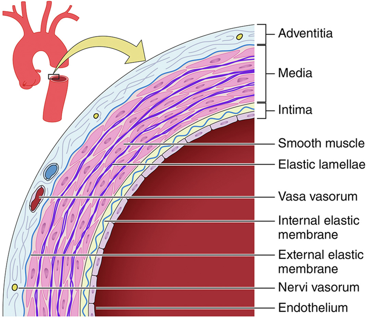
FIGURE 2. A Simplified Diagram Depicting the Key Histologic Components of the Aortic Wall.

The medial layer in human aortas contains >50 alternating layers of elastin and smooth muscle cells (whereas only 5 are shown in this simplified illustration). Adapted (cropped) from “Illustration of tunics of the arteries vs veins” by Malgosia Wilk-Blaszczak, used under CC-BY 4.0. “Illustration of tunics of the arteries vs veins” is adapted (cropped) from figure 20.3 in BC OpenStax Anatomy and Physiology used under CC-BY 4.0.
