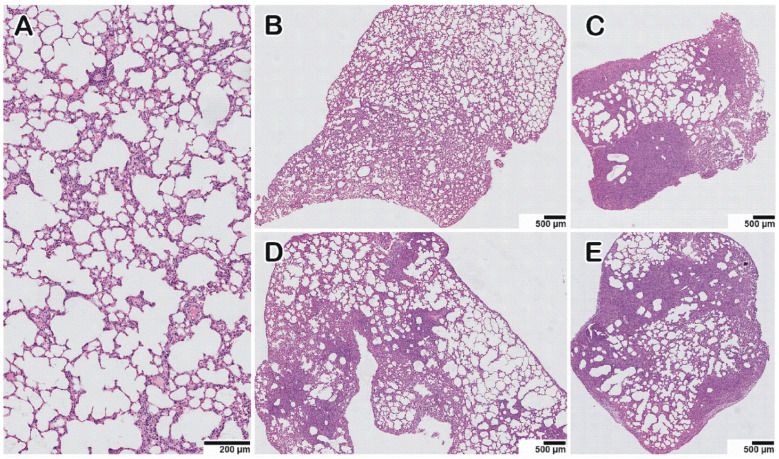
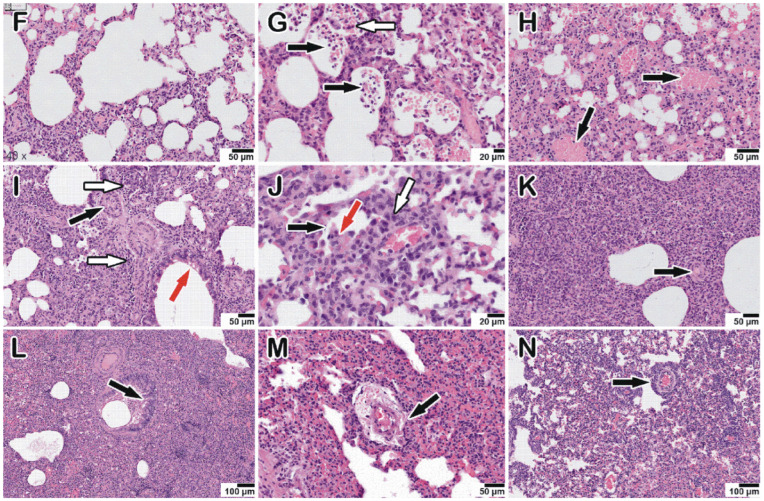
Figure 5.
Histopathological alterations in hamster lungs monoinfected (HAdV-5, SARS-CoV-2, IAV) and coinfected with HAdV-5 and SARS-CoV-2. (A) mock-infected control animal, normal lung. (B–E) Representative images showing pathological changes in lung tissue after infection: (B)—with HAdV-5; (C)—with HAdV-5 and SARS-CoV-2 simultaneously; (D)—with SARS-CoV-2; (E)—with HAdV-5 and, 3 days later, with SARS-CoV-2. In both cases of coinfection with two viruses (C,E), there was a significant increase in the consolidation zones and pronounced perifocal compensatory atelectasis. (F) Manifestations of adenovirus infection: edema and mixed inflammatory cell infiltration of the interalveolar septa and, in the upper part of the image, pronounced plethora of the capillaries. (G–K) Typical pathological changes found in mixed infections. (G) Blood (black arrows), macrophages, and syncytium (white arrow) in the lumen of the alveoli and the activation and hyperplasia of type II pneumocytes. (H) Hemorrhage in the alveoli (black arrows). (I) Spasm of small vessels of the arterial type (black arrow); perivascular edema and perivascular lymphocytic infiltration (white arrow); desquamation of the epithelial lining of the bronchi (red arrow). (J) Destruction of the small bronchial wall (bronchiolitis, black arrow); desquamated bronchial epithelium in the lumen (red arrow); peribronchial and perivascular infiltration (white arrow). (K) Zone of consolidation of lung tissue with spasmodic arterioles (black arrow). (L–N) Manifestations of IAV infection: epithelial cell degeneration, vasculitis, and perivascular lymphocyte infiltration, respectively (black arrows).


