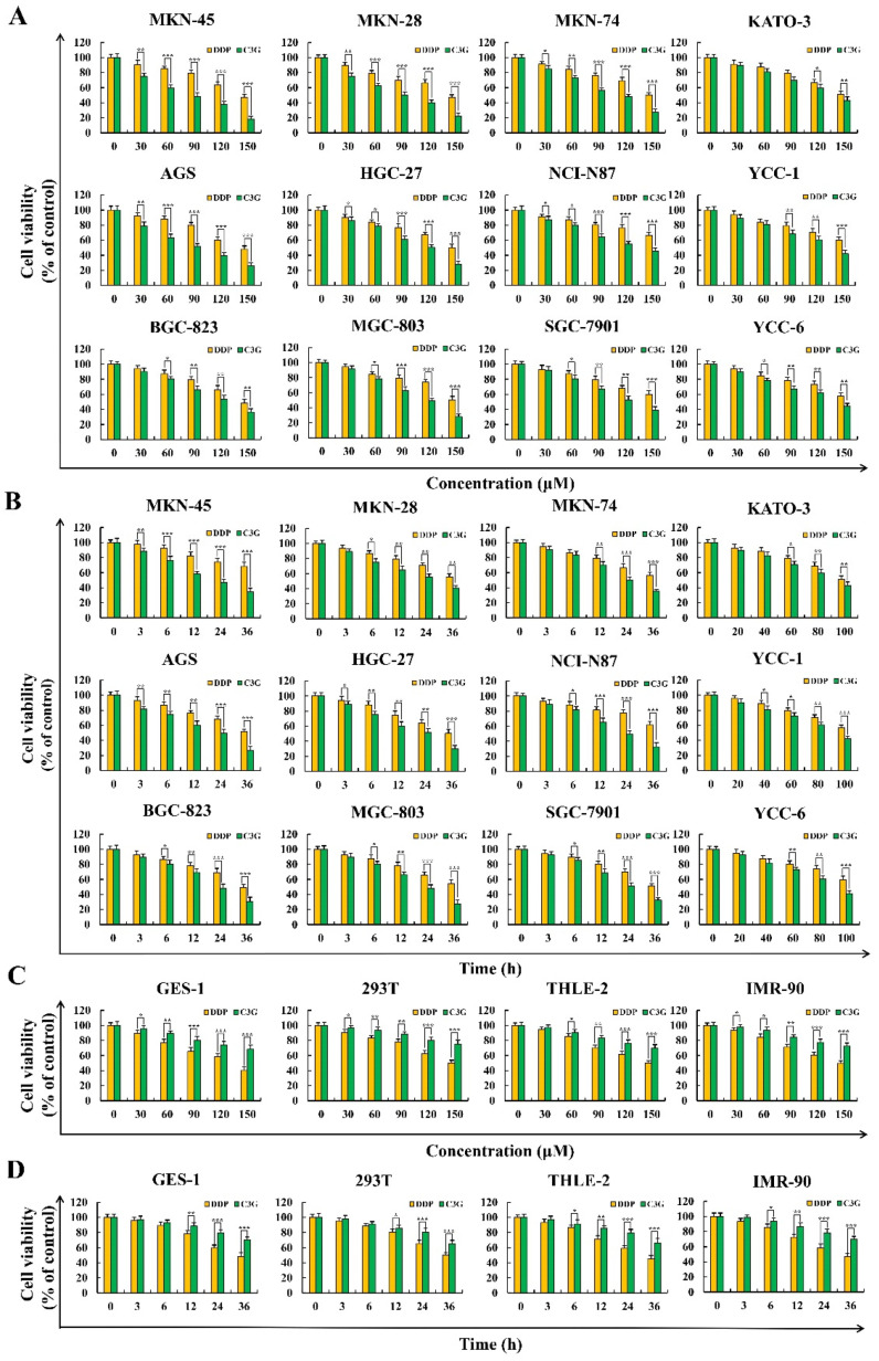
Figure 1.
Cytotoxic effects of C3G and DDP were detected using a CCK-8 assay. (A) Various concentrations (30, 60, 90, 120, and 150 µM) of C3G and DDP on the toxicity of 12 human GC cell lines. (B) Various time points (3, 6, 12, 24, and 36 h) of C3G and DDP treatment on the toxicity of 12 human GC cell lines. (C) Various concentrations (30, 60, 90, 120, and 150 µM) of C3G and DDP on the toxicity of four normal cells. (D) Various times point (3, 6, 12, 24, and 36 h) of C3G and DDP on the toxicity of four normal cells (* p ≤ 0.05, ** p ≤ 0.01, *** p ≤ 0.001 vs. DDP group).

