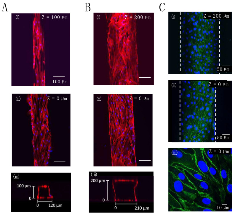
Figure 4.
Confocal microscopy images of stained cells present inside the microchannel of the gelatin device: (A,B) F-actin (red) and nuclei (blue) staining of NHDFs adhered at the top (i) or bottom (ii) of a microchannel, and confocal microscopy image of the cross-section of a microchannel (iii), the difference between (A,B) is the channel width and height; all other conditions were the same; (C) VE-cadherin (green) and nuclei (blue) staining of HUVECs adhered at the top (i) or bottom (ii,iii) of a microchannel. The dimensions of the microchannel were 100 μm width × 10 mm length × 100 μm height for (A) or 200 μm width × 10 mm length × 200 μm height for (B,C).

