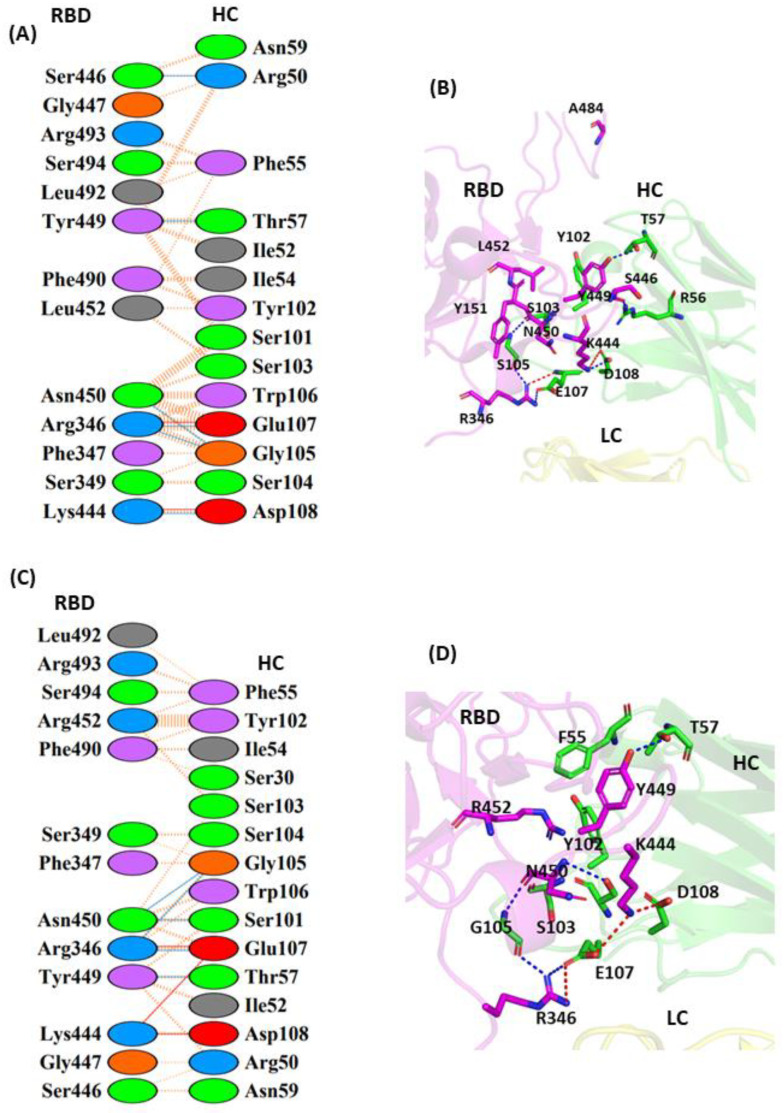
Figure 6.
Protein–protein interaction between the RBD and JMB-2002 monoclonal antibody: (A) 2D interaction diagram of binding interface of WT-RBD with JMB-2002 antibody; (B) 3D cartoon representation of intermolecular interaction of L452 residue of RBD with HC of JMB2002 antibody; (C) 2D interaction diagram of binding surface of L452R-RBD with JMB2002 antibody; (D) 3D cartoon representation of intermolecular interaction of L452R residue of RBD with HC of JMB2002 antibody. The interacting residues are shown in sticks. Only intermolecular interactions are indicated. RBD represents receptor-binding domain, HC represents heavy chain of monoclonal antibody, and LC represents light chain of monoclonal antibody.

