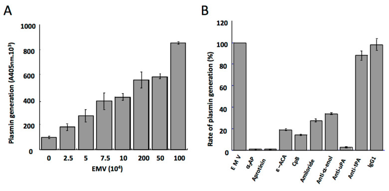
Figure 5.
Plasmin generation on membrane microvesicles derived from the human microvascular endothelial cell line HMEC-1. (A) Varying concentrations of endothelial microvesicles (EMV) were incubated with a fixed amount of plasminogen. Plasmin generation was detected with a chromogenic substrate. The amount of plasmin formed is a function of the concentration of microvesicles, i.e., of the amount of plasminogen activator (uPA) present at their membrane. (B) The specificity and characteristics of the activation of plasminogen at the microvesicle surface. Plasmin formed on the endothelial microvesicles is inhibited by a2-antiplasmin (α2-AP) and aprotinin. Binding and activation of plasminogen on endothelial microvesicles is prevented by the lysine analogue ε-aminocaproic acid (ε-ACA), carboxypeptidase B (CpB), and an anti-α-enolase (anti-α-enol) polyclonal antibody. The activity of uPA on the microvesicles is inhibited by amiloride and a specific polyclonal antibody anti-uPA, whereas an antibody anti-tPA or non-immune IgG has no effect [29].

