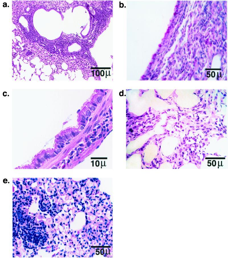
FIG. 2.
Representative lung sections of Cftr−/− mice after repeated exposure to B. cepacia isolate BC7. (a, c, d) Hematoxylin- and eosin-stained sections; (b) PAS/Alcian blue-stained section; (e) section stained with Giemsa. The histological sections demonstrate hypertrophy of PAS-positive cells in bronchiolar epithelia, hypertrophy of Clara cells, mucus retention in airways, inflamed parenchyma characterized by marked hypertrophy of resident interstitial cells with infiltration by macrophages and neutrophils, and consolidation of alveolar airspaces by an exudation of inflammatory cells and debris.

