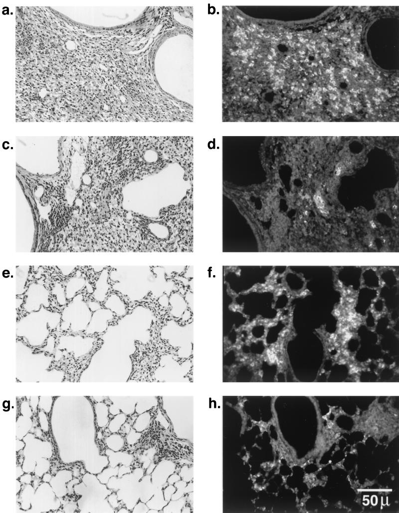
FIG. 3.
Immunolocalization of B. cepacia in the lungs of Cftr+/+ and Cftr−/− mice infected with isolate BC7. Paraffin sections (5 μm thick) were deparaffinized, rehydrated, heated in 10 mM sodium citrate buffer, pH 6.0, blocked with 5% normal donkey serum, and incubated overnight at 4°C with anti-B. cepacia antibody (diluted 1:1,000), and the bound antibody was detected by CY-3-conjugated anti-rabbit immunoglobulin G. (b and d) Bacteria in inflamed bronchoalveolar areas of Cftr−/− and Cftr+/+ mouse lungs, respectively; (f and h) bacteria in infiltrated and inflamed parenchyma of Cftr−/− and Cftr+/+ mouse lungs, respectively; (a, c, e, and g) hematoxylin- and eosin-stained sections corresponding to panels b, d, f, and h, respectively.

