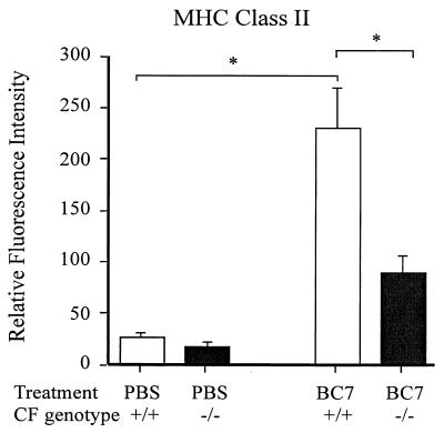
FIG. 6.
Surface expression of MHC-II molecules on alveolar macrophages recovered by BAL. Cells from 100 μl of BAL fluid were incubated with 20% FBS for 30 min, washed with 10% FBS, and incubated with fluorescein isothiocyanate-conjugated anti-CD11c (not shown) and phycoerythrin-conjugated anti-MHC-II. Cells were washed to remove excess antibodies and fixed with 1.6% paraformaldehyde, and fluorescence was quantified by flow cytometry. Macrophages were gated by a combination of light scattering and CD11c staining. The level of MHC-II expression was assessed on the gated cells. Asterisk, statistically significant difference between the groups, as determined by ANOVA with correction for multiple comparisons (Sheffe).

