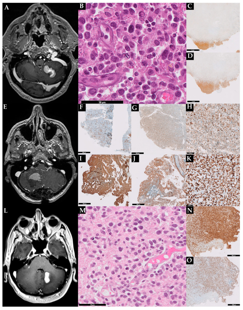
Figure 1.
Radiological and histopathological features of the three described cases. Case 1: brain MRI showing a mass in the left cerebellar hemisphere (A); hematoxylin and eosin with sheets of large pleomorphic cells (B); immunoreactive for CD20 (C); the lymphoma exhibited a high proliferation index (70%) (D). In (C,D) is also evident a large area of necrosis. Case 2: MRI showing a mass in the right hemisphere (E). Histopathological analisys revealed sparse CD3+ (F) and Bcl2+ (G) T-cells and a B-cell lymphoproliferation expressing MUM1 (H), CD20 (I), and p53 (J); the lymphoma exhibited a high proliferation index (K). Case 3: brain MRI showing a mass in the right cerebellar hemisphere close to the dentate nucleus (L); cerebellar sample staining in HE large lymphoid cells with centralistic morphology and perivascular preservation (M); the lymphoma expressed PAX5 (N) and exhibited a high proliferation index (O).

