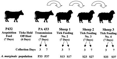
FIG. 1.
Experimental design. Calf PA432 was inoculated with 106 ml of infected blood (Virginia isolate of A. marginale) from PA431 (parasitemia = 0.9%) and served as the donor for infection of D. variabilis males. Calf PA432 was infested with 781 male D. variabilis ticks that were placed in orthopedic stockinettes attached to the calf when the ascending parasitemia was 4.4%. The ticks were allowed to feed for 7 days, after which they were removed and held in a humidity chamber for 6 days. The ticks were then allowed to feed on calf PA433 for 7 days, and then they were transferred directly and successively to feed for 7 days on sheep 1, 2, and 3. Forty ticks were removed from each host (PA433 and sheep 1, 2, and 3) on days 3 and 7 of tick feeding. The ticks were dissected, and salivary glands from the groups of 20 ticks were pooled and used for msp2 expression site cloning and sequence analysis.

