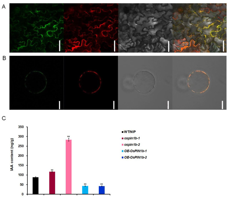
Figure 4.
Subcellular localization of OsPIN1b and auxin contents in lamina joints. (A,B) 35S:OsPIN1b-sGFP fusion construct and plasma membrane marker pm-rbCD3-1008 were simultaneously co-expressed in Nicotiana benthamiana epidermal cells (upper) and rice protoplasts (lower). From left to right, represents green fluorescence of 35S:OsPIN1b-sGFP, red fluorescence of membrane marker pm-rbCD3-1008, bright-field images, and yellow merged fluorescence, respectively. Scale bars = 10 μm. (C) Auxin contents in lamina joints of 7-day-old WT/NIP, ospin1b-1, ospin1b-2, OE-OsPIN1b-1, and OE-OsPIN1b-2. The three biological repeats were used in these experiments. The data are mean ± SD (n = 3) and asterisks indicate the significant differences in the above-mentioned lines (** p < 0.01; Student’s t-test).

