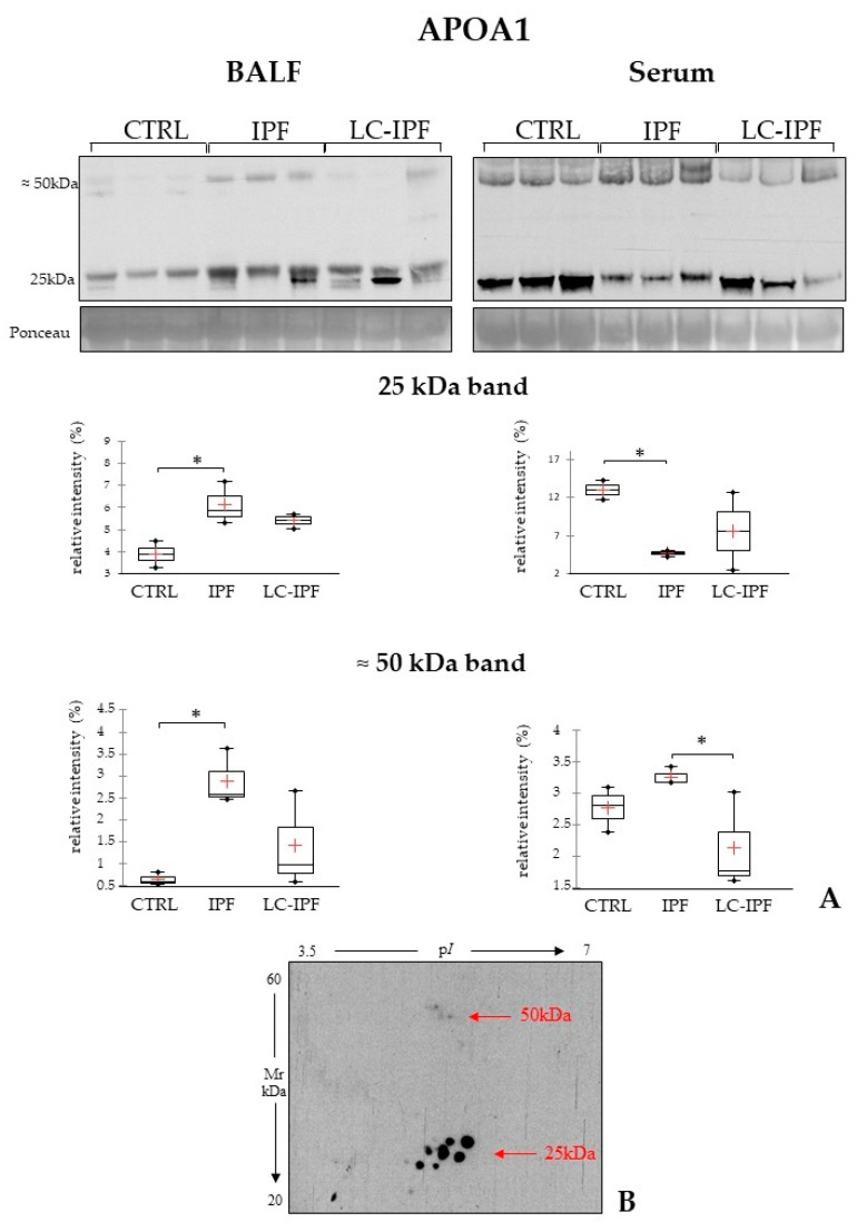
Figure 8.
(A). Western blot analysis of APOA1 in BALF and serum samples. Statistical analysis was performed by Kruskal–Wallis test and Dunn correction and reported in the Box plots (∗ p-value < 0.05). Ponceau red is reported to assure the equal loaded amount of sample and for the intensity normalization. BALF and serum samples used for WB were three for CTRL, three for IPF, and three for LC-IPF. (B). In the bottom, the two-dimensional Western blot of APOA1 in an IPF BAL sample is reported. Classical protein species of APOA1 are highlighted at 25 kDa and other protein species at higher molecular weight (50 kDa) are also evidenced in IPF.

