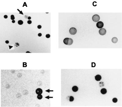
FIG. 3.
Reciprocal phase variation of P120 and surface masking of P56. Panels A and B are colony blots of the two clonal populations shown, respectively, in Fig. 2A and B that were immunostained with MAb 26.7D to P120. Phase variants in the predominant clonal populations are indicated by arrows. A sectored colony is indicated by the arrowhead in panel A. Panels C and D are replicate colony blots made from the same plate of a clonal population of 1630 that were immunostained, respectively, with anti-P56 Ab (C) or MAb 26.7D (D), showing the reciprocal staining patterns obtained with these two Abs.

