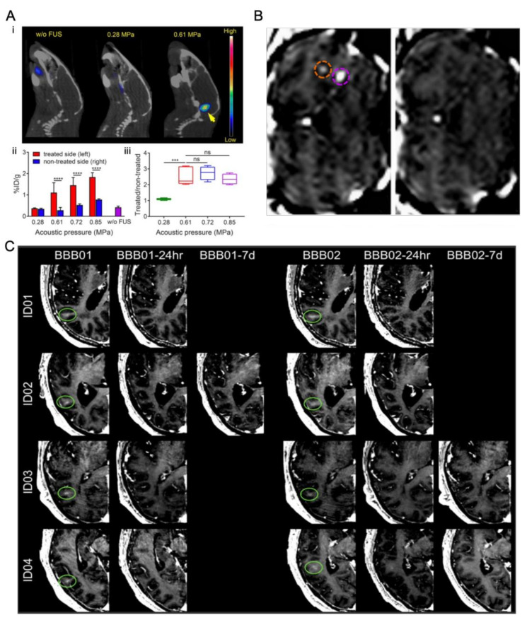
Figure 7.
Representative studies for BBB opening by ultrasound-driven microbubbles in vivo. (A) (i) In vivo PET/CT images of 64Cu-CuNCs without focused ultrasound (FUS) treatment and under 0.28 and 0.61 MPa FUS pressure in WT mice at 24 h post intravenous injection. (ii) Quantitative analysis of 64Cu-CuNC uptake in FUS-treated left pons and nontreated right pons of WT mice under 0.28, 0.61, 0.72, and 0.85 MPa FUS pressures as well as in the pons without FUS treatment in WT mice. (iii) Uptake ratios of the treated sites vs. nontreated sites of the FUS-treated mice under different pressures (*** p < 0.001, **** p < 0.0001, n = 4−5) [13]. (B) Coronal MR image after intravenous injection of gadodiamide 1 h after sonication, showing the presence of gadodiamide in the brain parenchyma confirming the local disruption of BBB. Two opening sites under different ultrasound treatment are circled (purple circle 0.3 MPa and orange circle 0.45 MPa). 6 days after sonication, coronal MR image confirming that the BBB is closed and the procedure is reversible [113]. (C) Brain CT images (gadolinium enhancement in T1-weighted) of patients 1–4 immediately after the BBB opening procedure (BBB01). The BBB opening was closed after 24 h (stage 1) in patients 1, 3, and 4 (BBB01-24 h) and in patient 2 on the seventh day of MRI follow-up (BBB01-7d). For stage 2 treatment, the BBB was closed in patients 1 and 2 after 24 h and in patients 3 and 4 in the following MRI study on day 7 (BBB02-7d) [116].

