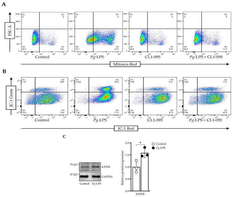
Figure 2.
P. gingivalis-LPS induced oxidative stress and decreased membrane potential. (A) Cells were stained with MitoSOX Red, and ROS-producing cells were sorted using flow cytometry. (B) JC-1 staining was used to assess the decline in the membrane potential. (C) Western blot analysis of 4-HNE and GAPDH was used as a loading control (n = 3). * p < 0.05.

