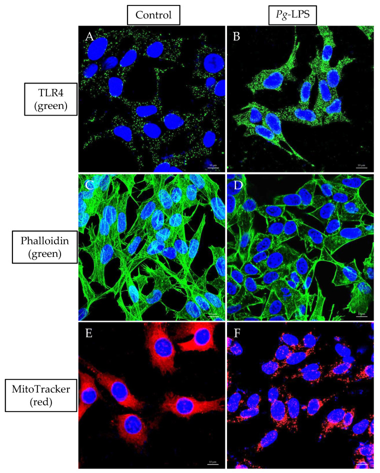
Figure 7.
Immunolabeling of TLR4, Actin and MitoTracker. The cells were incubated with Anti-TLR4 Antibody (green), phalloidin (green) and MitoTracker (red) conjugated dyes. DAPI (blue) was used for nuclear counterstaining. Untreated (A,C,E) and LPS-treated (B,D,F). A 63× oil objective was used; scale bars indicate 10 μm.

