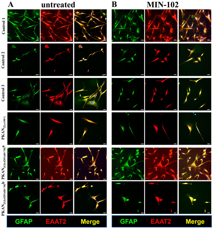
Figure 1.
Characterization of astrocytes (50–60 days of differentiation). Controls and PKAN patients’ astrocytes were differentiated from NPC by growing them in astrocytes’ medium in the presence (B) or absence (A) of MIN-102 (100 nM). Cells were then fixed and stained with the astrocyte markers GFAP (green) and EAAT2 (red) to identify mature astrocytes. Nuclei were stained with Hoechst (blue). Images were taken on Zeiss Axio Observer.Z1 equipped with Hamamatsu 9100-02 EM CCD camera using 20× objective. Scale bar = 20 μm.

