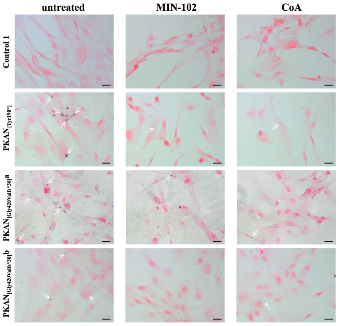
Figure 5.
Iron accumulation in astrocytes (80 days of differentiation). Astrocytes (Control 1 and PKAN patients) were differentiated in the presence or absence of 100 nM MIN-102 or 25 μM CoA. Fixed astrocytes were stained with Perls reaction to detect iron granules (blue) and counterstained with nuclear fast red. Images were taken on Zeiss AxioImager M2m equipped with AxioCam MRc5 using 40× objective. White arrows indicate iron granules. Scale bar = 20 μm.

