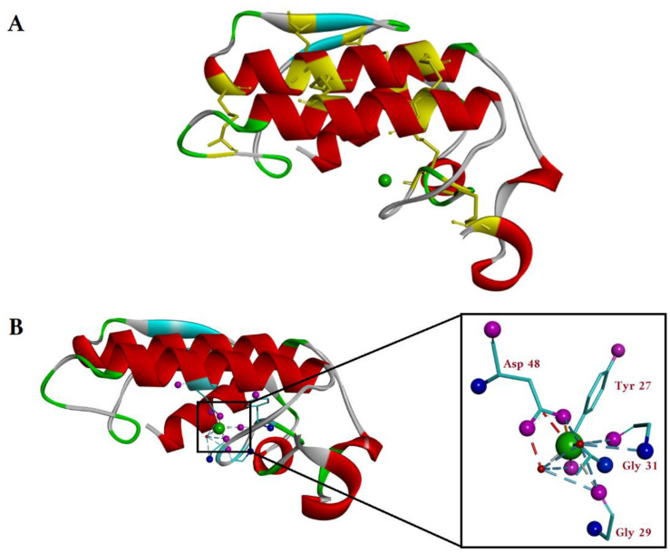
Figure 3.
(A) Top view of the crystal structure of G-IA sPLA2 (Naja naja) (PDB-1PSH). The α-helices and β-sheets are shown in red and light blue. The cysteines forming the seven disulfide bonds are shown in yellow, also marking in yellow their position on the protein backbone. The calcium ion is shown as a green sphere in the enzyme active site. (B) Side view of the crystal structure of the sPLA2 with the Ca2+ coordination shown in detail in the zoomed insert. The oxygen atoms and the nitrogen atoms of the amino acids coordinated with Ca2+ (green sphere) are colored pink and blue, respectively. The Ca2+ is directly coordinated with carboxy group from Asp48 of one α-helix and by the C=O backbone groups of Tyr27, Gly29 and Gly31 from the opposite loop. (Image generated using BIOVIA Discovery studio.)

