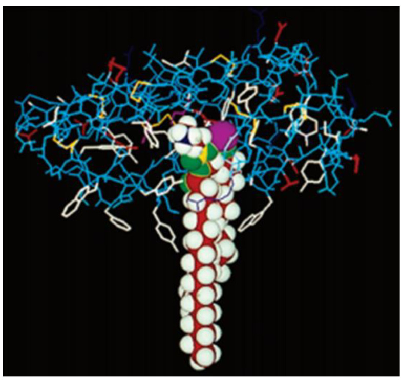
Figure 5.
Cartoon depicting the X-ray crystal structure of cobra venom sPLA2 (wireframe) with dimyristoyl phosphatidylethanolamine (shown in a space-filling model) bound to it. The active site dyad His48/Asp99 and the Ca2+ ion are depicted together as a purple sphere. The aromatic interfacial residues Phe-5, Trp-19, Tyr-52 and Tyr-69 of PLA2 are shown in white. These residues of the enzyme are reaching as deep as 9–10 carbons in the acyl chain of the fatty acid while the rest of the chain is submerged into the lipid bilayer. Reprinted with permission from [21]. Copyright 1994, Elsevier.

