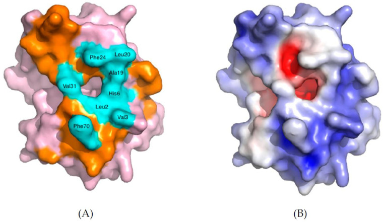
Figure 8.
Space-filling model of human group IIA sPLA2 (PDB—3U8B). (A) The amino acids which form the i-face are colored orange, and the ones which create the entrance are highlighted in cyan blue, while the rest of the protein is shown in pink. (B) The electrostatic charge distribution is depicted either in blue (positive charge) or in red (negative charge). The white areas charge neutrality (non-polar), which corresponds to the hydrophobic entrance of the left figure. Reprinted from [26].

