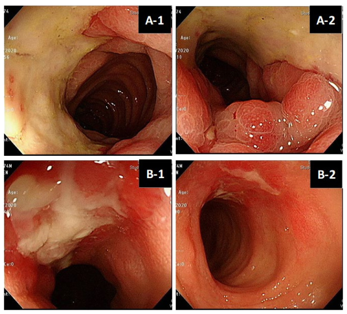
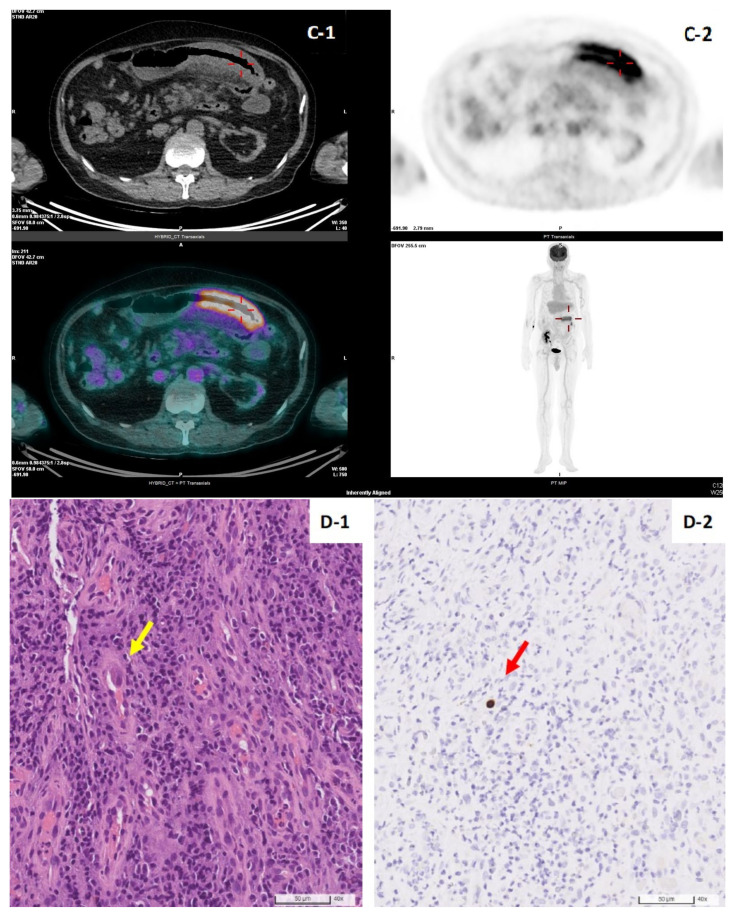
Figure 2.
(A,B). Colonoscopy features of living donor liver transplant patient No.1. Well-demarcated longitudinal ulceration (around 3 cm in diameter) in descending colon with colonic mucosa edematous change and bowel wall thickening, causing intra-luminal narrowing (A-1,A-2). Remission of ulceration 2 weeks later after antiviral therapy follow-up (B-1,B-2). (C). Positron Emission Tomography/Computed Tomography (PET/CT) of patient No.1. PET/CT illustrated segmental colon wall thickening at distal T-colon and proximal D-colon by CT scan (C-1, arrow) and increased FDG uptake in the colon wall, SUV max:7.6 by PET (C-2, arrow), respectively. (D). Histopathology of CMV colitis in patient No.1. Histological hematoxylin and eosin staining (×40 objective) detection of CMV inclusion bodies (owl’s eye) (yellow-arrow), biopsy specimen of an ulcer at descending colon (D-1). Positive CMV immunohistochemistry (IHC) staining (×40 objective) (red-arrow) (D-2).


