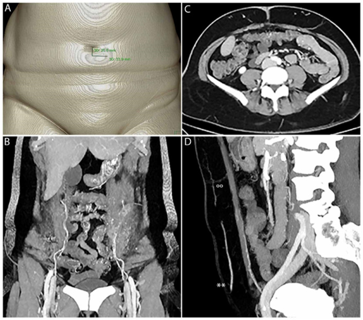
Figure 1.
CTA enables fast 3D PM with clear delineation of SP, P, F, M and the IEA. SP must be measured manually in relation to the umbilicus (A). (B) shows PM in 3D. The origin of the IEA, including M with type II branching on the left and type I branching on the right, is clearly visible. In (C), F and P of the perforators on the left and right side are clearly visible. IEA, M, F, P(°°) and SP can be clearly seen within the sagittal view (D), including the superficial epigastric artery (SIEA**).

