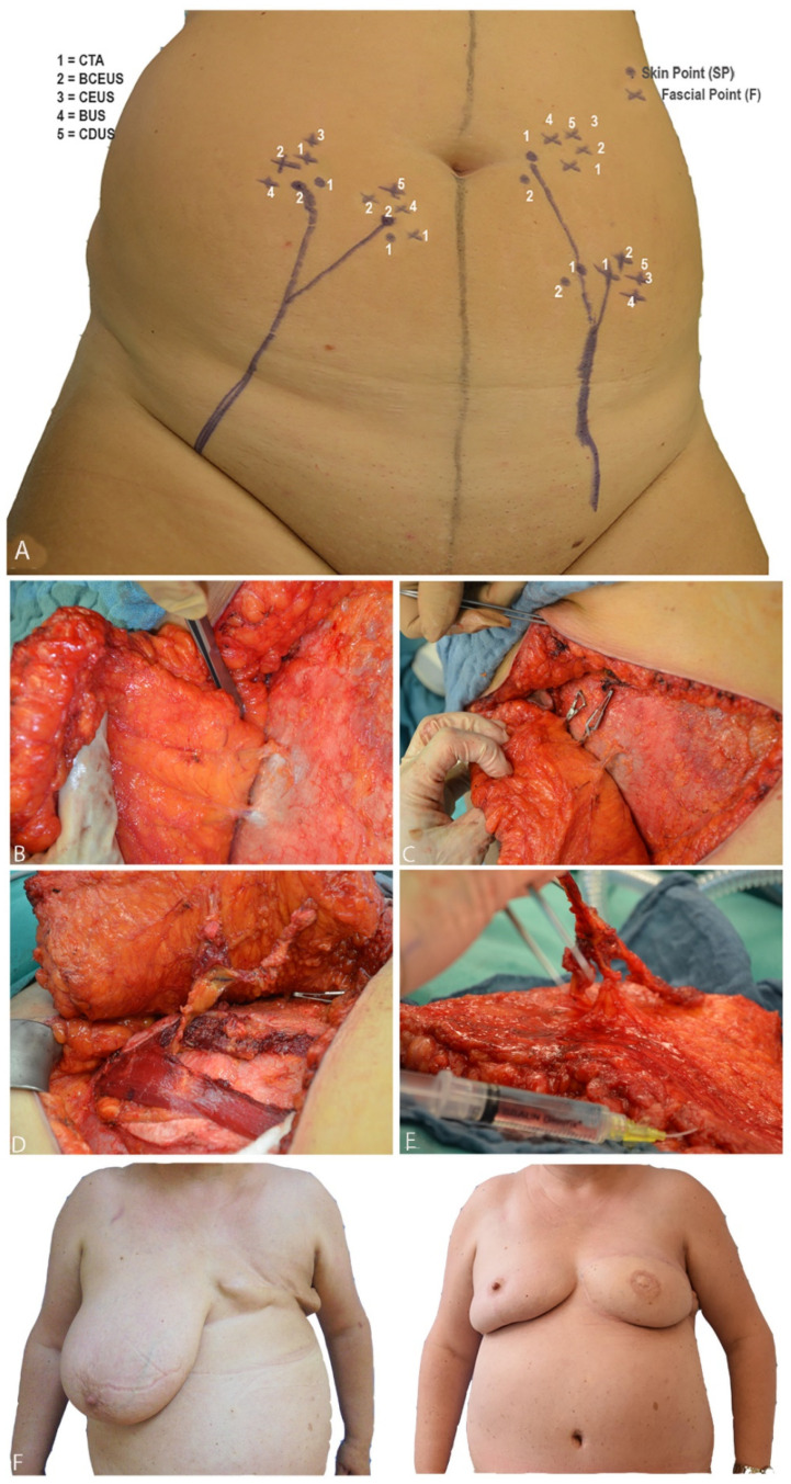
Figure 4.
(A) shows preoperative PM. When BCEUS, BUS, CEUS and CDUS were used, landmarks could be directly navigated on the abdomen. (B) illustrates F and P of the respective perforators. In (C), the clamping test is shown. (D) portrays the intramuscular dissection of the perforator vessels down to the origin of the DIEA. (E) shows the harvested fasciocutaneous DIEP flap, including the perforator vessels. (F) shows pre- and postoperative pictures of a patient after breast amputation and consecutive DIEP flap reconstruction, including contralateral breast reduction.

