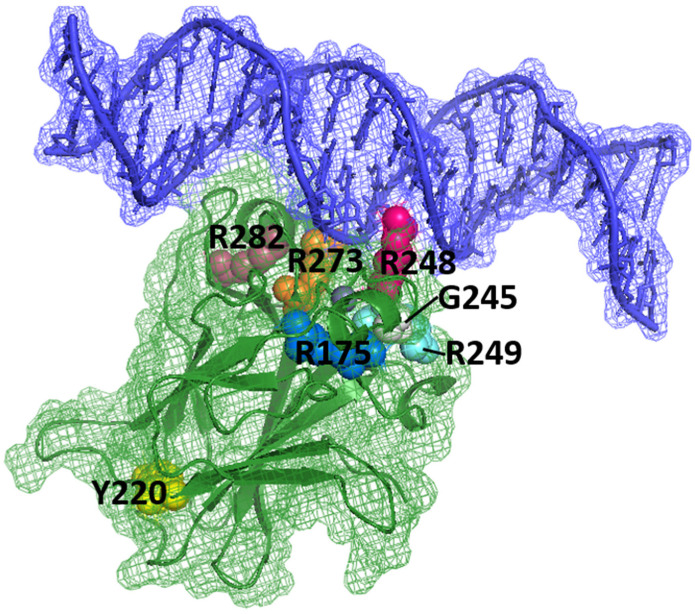
Figure 1.
p53 DBD (green) in complex with DNA (blue). The locations of amino acids R175, Y220, G245, R248, R249, R273 and R282, which are sites of key carcinogenic mutations (“hot spots”), are shown (PDB identifier: 1TSR) [4]. DNA is in direct interaction with R273 and R248, and several “hot spots” are present near this interaction (R249, G245, R282). Other “hot spots” are far from the surface involved in DNA binding, for example, Y220.

