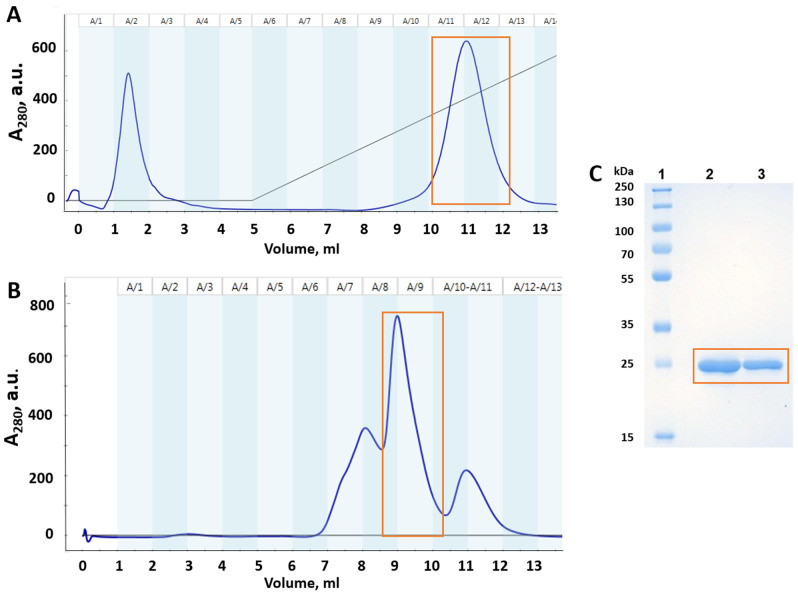
Figure 2.
Purification of mutant p53(Y220C) DBD. Orange square shows the purified mutant p53(Y220C) DBD protein: (A) heparin-sepharose chromatography profile (dark blue line—absorbance at 280 nm); (B) gel filtration chromatography profile; (C) SDS-PAGE monitoring of mutant p53(Y220C) DBD after gel filtration chromatography: Lane 1—Protein molecular-weight marker; Lane 2—p53(Y220C) DBD (10 µg/well); Lane 3—p53(Y220C) DBD (5 µg/well).

