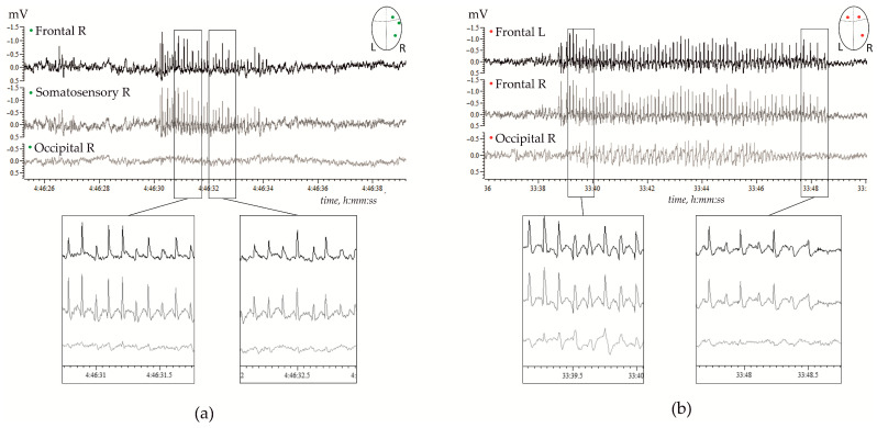
Figure 1.
Examples of spontaneous spike-wave discharges (SWDs) in electrocorticograms recorded in freely moving adult WAG/Rij rats; (a)—11 months old subject, and (b)—7 months old subject. (a) Regular high-voltage 8–10 Hz SWDs were expressed in the right hemisphere (R) over the frontal and parietal (somatosensory) cortical areas, but were hardly seen over the occipital area. (b) High-voltage 8–10 Hz SWDs were bilaterally synchronized, and were present in the left (L) and right frontal cortical areas, and were seen in the occipital cortex.

