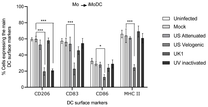
Figure 11.
Effect of EAV infection on MoDC differentiation. Monocytes were inoculated at MOI of 5 with different strains of live and UV inactivated virus. Cells were incubated for 2 h after infection prior to the addition of 1000 U/mL of GM-CSF and 500 U/mL of IL-4. Cultures were incubated further for 16 h, after which cells were harvested and stained to assess phenotype by flow cytometry. For the different treatments, the same number of events (10,000) were analyzed in the target gate. There were significant differences during differentiation in the phenotype of MoDC infected with the Velogenic strain. Results were represented as the percentage surface marker expression ± SEM (n = 6). A 2-way ANOVA with Bonferroni post-test was used to compare replicate means. * and *** indicate significant differences between sample means where p < 0.05 and 0.001.

