Figure 4.
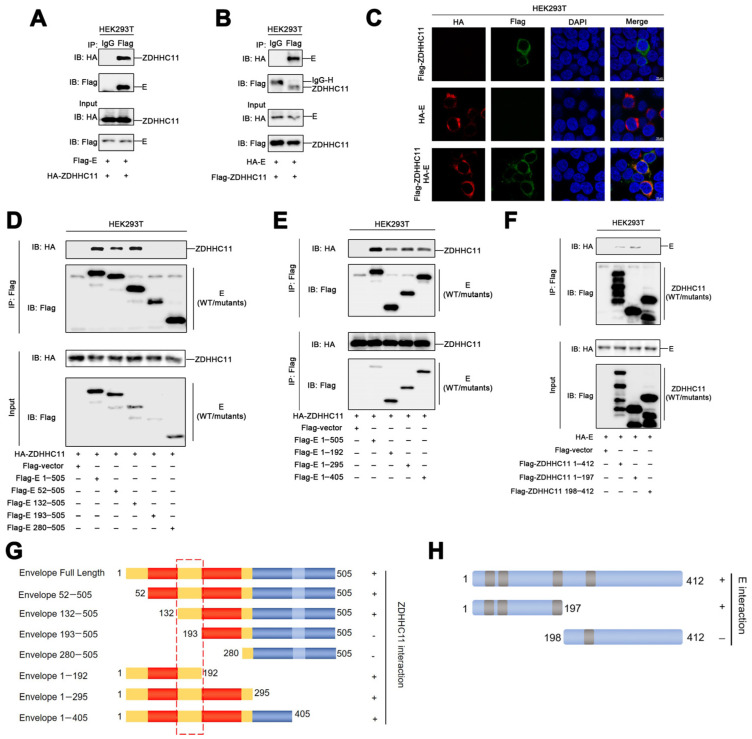
ZDHH11 interacts with the envelope protein of ZIKV. (A,B) HEK293T cells were transfected with plasmids encoding the envelope protein and ZDHHC11 for 24 h, lysed for co-immunoprecipitation assays with the indicated antibodies, and detected using immunoblotting with the indicated antibodies. (C) HEK293T cells were transfected with HA-E or Flag-ZDHHC11, or co-transfected with HA-E and Flag-ZDHHC11. The sub-cellular localizations of HA-E (red), Flag-ZDHHC11 (green), and the nuclear marker, DAPI (blue) were analyzed with CLSM. (D,E) HEK293T cells were transfected with plasmids encoding ZDHHC11 and the WT or truncated envelope proteins for 24 h, lysed for co-immunoprecipitation assays with the indicated antibodies, and detected using immunoblotting with the indicated antibodies. (F) HEK293T cells were transfected with plasmids encoding the envelope protein and ZDHHC11 or truncated ZDHHC11 for 24 h, lysed for co-immunoprecipitation assays with the indicated antibodies, and detected using immunoblotting with the indicated antibodies. (G) Diagrammatic representation of the full-length and truncated envelope protein. Domain I, II, and III are marked by yellow, red and blue. (H) Diagrammatic representation of the full-length and truncated ZDHHC11 enzyme.

