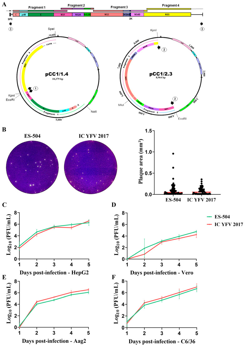
Figure 2.
Recovery and assessment of clone-derived YFV. (A) Strategy for the assembly of YFV genome. The genome was divided into 4 fragments and reassembled into two main plasmids bearing the extremities (pCC1/1.4) and the central part (pCC1/2.3) of the viral cDNA. Black arrows 1 to 3 represent the primer pairs used in the amplification rounds before in vitro transcription of the template cDNA as described in Section 2.2 and Supplementary Table S1. Arrows numbered 1 represent the primer pair used to amplify the entire plasmid pCC1/1.4, arrows numbered 2, the primer pair employed in the amplification of the central part of the genome (fragment 2.3), and arrows numbered 3, are the primers used to amplify the complete viral cDNA. (B) Plaque morphology of parental YFV ES-504 and clone-derived virus YFV_2017. Plaque areas were measured in ImageJ software, and the results were plotted and compared in GraphPad Prism 8 with the Mann–Whitney test. (C–F) Growth curves in different cell lines: HepG2 (C), Vero (D), Aag2 (E), and C6/36 (F). The viral titers were transformed in log10 and plotted in GraphPad Prism 8. Statistical analyses were applied to each time point using the unpaired t-test to compare viral titers of YFV ES-504 and YFV_2017.

