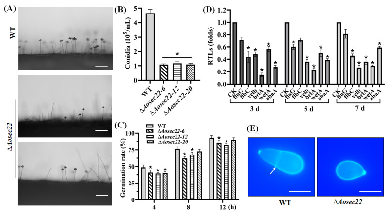
Figure 2.
Role of AoSec22 in conidiation and conidial morphology. (A) Microscopic images (scale: 50 μm) for conidiation of the WT and mutant strains on PDA. (B) Spore yields assessed from 15-days-old cultures. (C) Spore germination rate during normal incubation. (D) Relative transcription levels (RTLs) of sporulation-related genes in the WT and mutant strains. An asterisk (B–D) indicates a significant difference between the mutant and WT strains (p < 0.05). CK was used as the standard (RTL = 1) for statistical analysis of the RTL of each gene under a given condition. (E) Microscopic images (scale: 10 μm) of conidia stained with calcofluor white (CFW). The white arrow indicates the septum.

