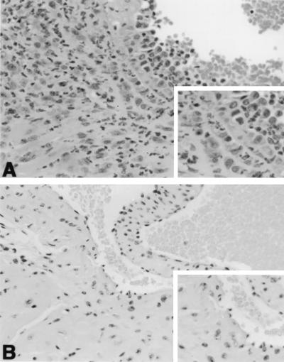
FIG. 1.
Hematoxylin-eosin stain of representative heart tissues. (A) Heart base of a B6 TCR α−/− mouse at 45 days of infection. Note margination of leukocytes along the endothelial surface above the aortic valve, and infiltration of leukocytes in the wall and adventitia of the root of the aorta (upper left and insert) and ventricular myocardium (lower left). (A) Heart base of a B6 mouse at 45 days of infection. There is no inflammation remaining near the aortic valve (valve leaflet in center and insert) or root of the aorta (upper left). Magnification, ×175.

