Figure 3.
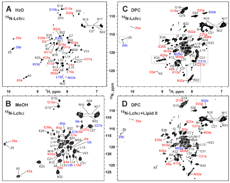
2D 15N-HSQC spectra and resonance assignment of Lchα in different environments. (A) H2O (Lchα 0.3 mM, pH 4.0, 30 °C). (B) d3-methanol (Lchα 0.3 mM, pH 3.5, 27 °C). (C) DPC micelles solution (Lchα 0.35 mM, DPC 30 mM, D:P = 86:1, pH 5.8, 45 °C). (D) DPC micelles solution with lipid II (Lchα 0.18 mM, lipid II 0.72 mM, DPC 42 mM, Lchα:lipid II:DPC = 1:4:240, pH 5.8, 45 °C). The resonance assignment is shown. The signals of the major and two minor forms of the peptide are marked in black, red, and blue, respectively. The signals of the minor forms are additionally marked with “a” or “b”. The residue names are given in the one letter code format, where 2,3-didehydroalanine, 2,3-didehydrobutyrine, lanthionine, and methyllanthionine are abbreviated as X, Z, U-C, and B-C, respectively. Areas highlighted by rectangles in panel (C) are discussed in Section 2.5.

