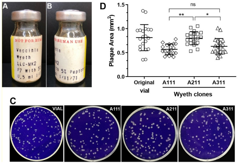
Figure 1.
Clone isolation of VACV-Wyeth and plaque phenotype. (A,B) Original vial of lyophilized VACV-Wyeth propagated in LLC-MK2 in 1971. (C) Representative images of crystal-violet stained BSC-40 cells infected with 10−6 dilution of the original vial before plaque selection and stocks of Wyeth clones A111, A211, and A311. (D) Random viral plaques (n = 20) were photographed at 4× magnification, and the individual areas were measured. Circles: plaques of the original vaccine vial; diamonds: plaques of clone A111; squares: plaques of clone A211; triangles: plaques of clone A311. Asterisks: * p ≤ 0.05 and ** p ≤ 0.01; ns means non-significant.

