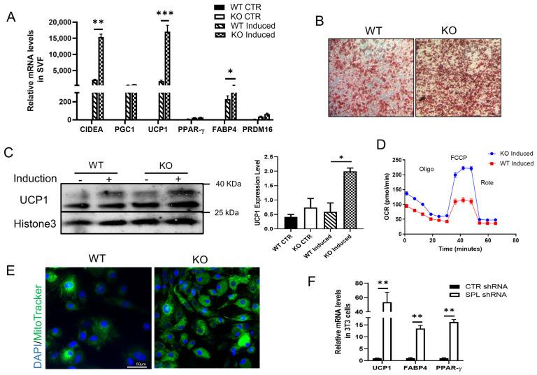
Figure 3.
In vitro browning induction showed increased browning gene expression in SVF-derived adipocytes from WT and KO mice. vWAT were harvested from WT and KO mice, cultured, and treated with a full browning induction medium to induce browning, and expression of markers was measured. (A) mRNA expression of CIDEA, PGC1, UCP1, PPAR-γ, and FABP4 relative to beta-actin in SPL KO and WT adipocytes. (B) Oil-red staining of SVF after full browning induced. (C) UCP1 protein expression level in vWAT cells from WT or KO mice after full browning induced. Relative protein expression of UCP1 compared to the expression of Histone 3 in vWAT induced. (D) Oxygen consumption rate (OCR) and (E) mitochondria tracker in WT and KO mitochondria function after browning induction. (F) Relative mRNA levels of UCP1, FABP4, and PPAR-γ in control or SPL shRNA transfected 3T3L1 cells after browning induction. Data were from at least three individual experiments. Two-way ANOVA with Tukey-Kramer post hoc test was used to compare the difference in (A,C). Unpaired Student’s t-test was performed to compare the difference between WT and KO in (F). * p < 0.05, ** p < 0.01, and *** p < 0.001. All data are presented as means ± SEM.

