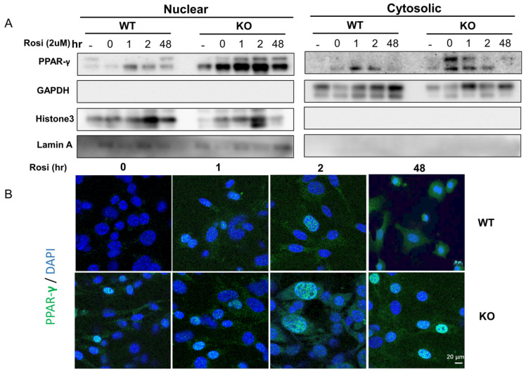
Figure 4.
SPL KO vWAT adipocytes promoted/retained more PPAR-γ in the nuclear after Rosiglitazone stimulation. Browning was induced in SVF vWAT adipocytes for 2 days and then treated with rosiglitazone at 2μM for another 48 h. (A) Western blot of PPAR-γ, GAPDH, Histone 3, and Lamin A in the nuclear fraction or cytosol fraction of vWAT harvested from WT and SPL KO mice before (0 h) and at 1 h, 2 h, and 48 h post-rosiglitazone treatment. (B). Immunofluorescent staining of PPAR-γ in WT and KO mice before (0 h) and 1 h, 2 h, and 48 h post Rosiglitazone treatment. Data were from at least three individual experiments. Scale bar = 20 μm.

