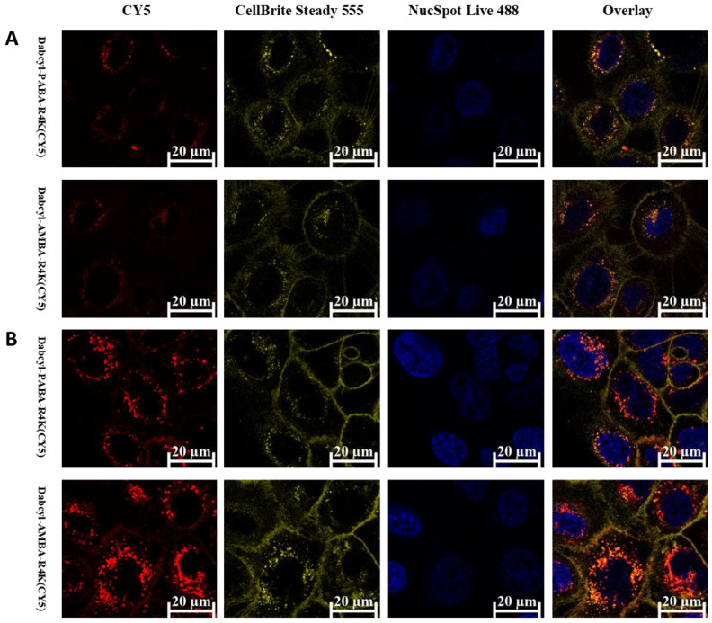
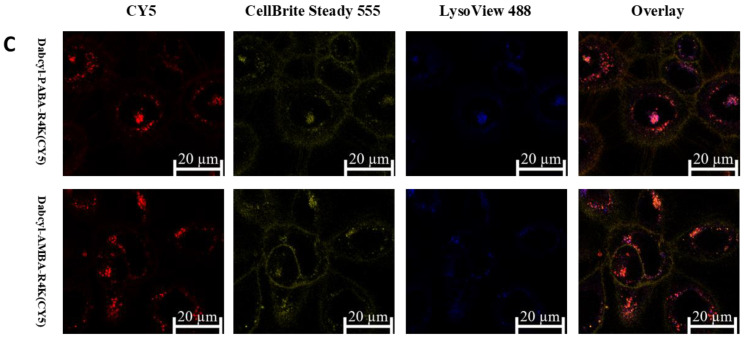
Figure 7.
Cellular distribution of peptides on SCC-25 cells visualized by confocal microscopy. Cells were incubated with the CY5-labelled peptides for 1hr (Red). CellBrite Steady was used to stain the membrane (green). Nuclei were stained with NucSpot Live 488 (Figure 6A,B blue), lysosomes were stained with LysoView 488 (Figure 6C blue). Concentrations of the peptides were (A) 5 µM, (B) 0.31 µM and (C) 5 µM in the three experiments. Imaging was performed by a Zeiss LSM 710 system.


