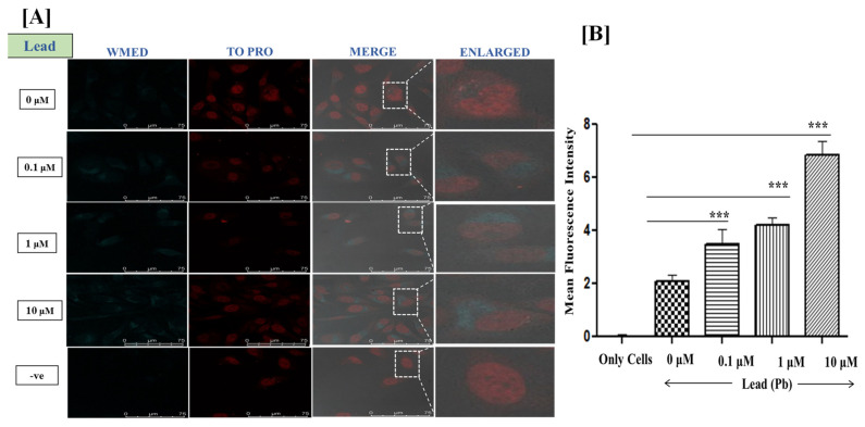
Figure 8.
(A) Confocal fluorescence microscopy images at 100× Magnification; Live HeLa cells were incubated with Lead ions at 0.1 μM, 1 μM, and 10 μM concentrations for 1.5 h. Staining of Live HeLa cells with 0.128 mg/mL WMED, images recorded for WMED at λex 405 nm/λem 450–550 nm. The Nucleus is stained with TO-PRO nuclear dye λex 641 nm/λem 661 nm; (B) quantification of lead ions using ImageJ software version 1.46r, *** p < 0.001.

