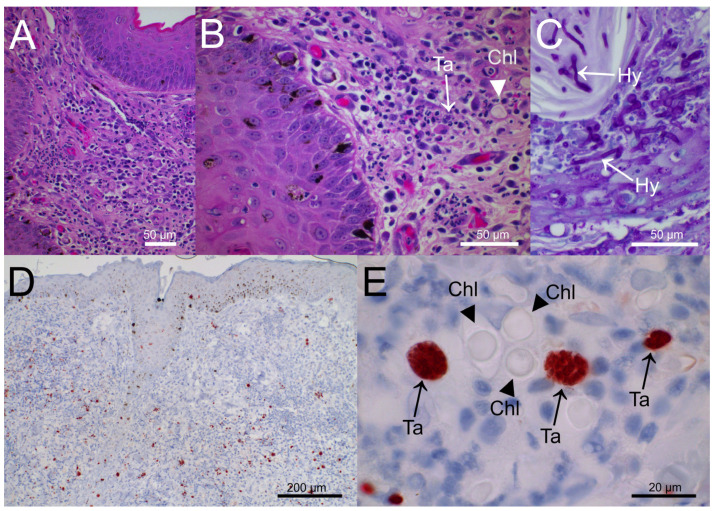
Figure 2.
Punch biopsy of the thorax nodule (A). Focally extensive pyogranulomatous inflammation of the upper dermis with scattered 10 µm diameter spheric microorganisms and moderately hyperplastic epidermis (HE stain). (B). Perivascular to diffuse pyogranulomatous dermatitis; few tachyzoites (Ta, arrow) and spherical structures evocative of fungal chlamydospores (Chl, arrowhead) are visible (HE stain). (C). Heavy colonization of hair (endothrix) and hair shaft by PAS-positive fungal filaments (hyphae, Hy, arrows). (D). Immunohistochemistry for Toxoplasma gondii, numerous immunolabelled (brown) protozoan tachyzoites scattered throughout dermis and epidermis. (E). Spheric fungal structures (chlamydospore-like elements, Chl, arrowheads) and immunolabelled (brown) parasitophorous vacuoles filled with tachyzoites (Ta, arrows).

