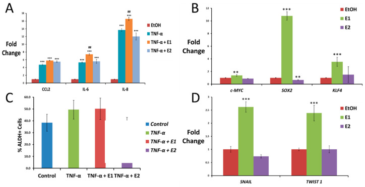
Figure 4.
Effect of TNF-α, estrone and estradiol on driving inflammation, stemness and EMT in HeLa cells. (A) Pro-inflammatory cytokine expression measured using qPCR in HeLa cells after treatment with 10 ng/mL TNF-α for 4 h alone or in combination with 10 nM E1 or E2 (data normalized to 1 for control (EtOH) using GAPDH as the housekeeping gene). (B) qPCR for the expression of the ES-TF c-MYC and SOX2 in HeLa cells after 3 weeks of exposure to 10 nM E1 or E2 (data normalized to 1 for control (EtOH) using GAPDH as the housekeeping gene). (C) Expression of ALDH activity measured using flow cytometry in HeLa cells untreated (control) or treated with 10 ng/mL TNF-α alone or in combination with 10 nM E1 or E2 for 3 weeks. (D) EMT transcription factor expression measured using qPCR in HeLa cells exposed to 10 nM E1 or E2 for 3 weeks (data normalized to 1 for control using GAPDH as the housekeeping gene). All data are graphed as mean ± SEM from experiments performed in triplicates and repeated at least 3 times. ** p < 0.01 *** p < 0.001 vs control; ## p < 0.05 vs TNF-α.

