Figure 1.
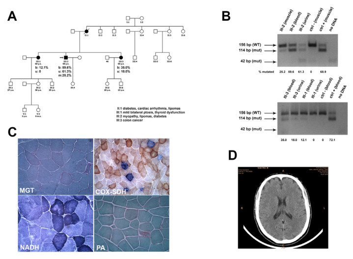
(A) Pedigree of family. (B) PCR-RLFP analysis of amplicons obtained from muscle, urine, and blood of the indicated subjects, electrophoresed on 4% agarose gel after BglI cut. The numbers under each lane indicate the estimated mutational load in the sample. (C) Morphological examination showed some RRFs at MGT, several COX-negative fibers, many of which are intensely SDH-positive (RRFs). Scale bar 50 μm. (D) Brain CT scan.

