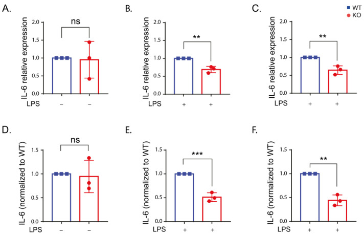
Figure 4.
IL-6 expression is reduced in the absence of LRRK2 after LPS stimulation. (A–C) IL-6 expression in the WT and KO cells under control conditions (A) and after LPS stimulation for 6 (B) and 24 h (C) using reverse transcription quantitative real-time PCR using β-actin and ribosomal protein L13A (RPL13A) as housekeeping genes. (D–F) The amount of IL-6 in the supernatant of the WT and KO cells under control conditions (D) and after LPS stimulation for 6 (E) and 24 h (F) using ELISA assay. The experiment was performed 3 times and t-test was used to compare the average IL-6 expression or level normalized to the corresponding WT. Error bar represents mean ± SD. p-values indicating statistically significant differences between the mean values are defined as follows: ns—not significant, ** p < 0.01, *** p < 0.001.

