Figure 7.
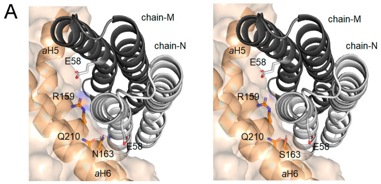
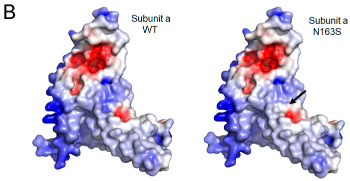
The impact of N163S mutation in the structural model of the mouse ATP synthase subunit a: (A) detail of the interface view between the c-ring and a subunit for WT (left panel) and p.N163S (right panel). Chain-N and chain-M of c-ring are displayed with cartoon representation (light and dark gray, respectively). Subunit a is displayed as surface with cartoon representation of the secondary structure elements, helices aH5 and aH6, implicated in the interaction. Relevant residues are represented as sticks and CPK colored; and (B) impact of p.N163S mutation on the electrostatic surface potential (ESP) of mouse subunit a. ESP for wild-type (left panel)) and its N163S (right panel) variant was calculated at pH 7.4 and 150 mM of salt using the APBS-PDB2PQR software suite (https://www.poissonboltzmann.org/ (accessed on 17 November 2022)) and then plotted using PyMOL. Position of N163S mutation is indicated by a black arrow. The range of change potential is −5 (red) to +5 (blue). The structural model of mouse subunit a (AF-P00848) was obtained from AlphaFold protein structure database [66,67].


