Figure 8.
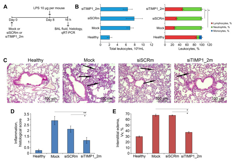
siTIMP1_2m effectively suppresses LPS-induced lung inflammation in vivo. (A) Experimental setup. Acute lung injury (ALI) was induced in mice (n = 6) by intranasal (i.n.) instillations of LPS (10 µg per mouse). Mice were pretreated with siTIMP1_2m administered i.n. 8 days before ALI induction. Mock and siSCR were used as controls. Mice were sacrificed 16 h after LPS challenge followed by collection of bronchoalveolar lavage (BAL) fluid and the lung tissue for subsequent analysis. (B) Total (left) and differential (right) leukocyte counts in the BAL fluid of healthy and LPS-challenged mice without treatment and after siTIMP1 administration. Four BAL samples from each experimental group were analyzed. (C) Representative histological images of lung tissue of ALI mice without treatment and after siTIMP1_2m administration. Haematoxylin and eosin staining. Original magnification ×200. Black arrows indicate inflammatory infiltration in the lung tissue. (D,E) The intensity of inflammatory infiltration quantified by the histological scoring system (D) and the volume density of interstitial edema (E) in the lung tissue of LPS-challenged mice without treatment and after siTIMP1 administration. The inflammatory scores and interstitial edema were calculated in 5 random fields in each lung sample, forming 30 random fields from each experimental group. Data are presented as mean ± standard deviation. * p ≤ 0.05, ** p ≤ 0.01, Mann–Whitney U-test.

