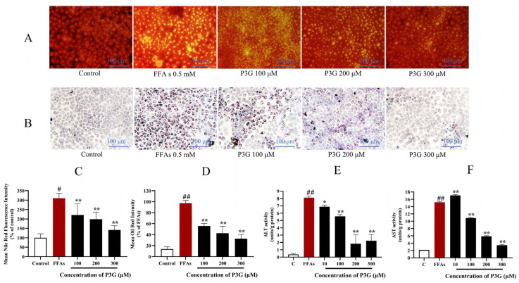
Figure 3.
Through the use of Nile Red and Oil Red O staining, the effects of purple corn anthocyanins (P3G) on FFA-induced lipid formation in L02 cells are demonstrated. (A) Nile Red staining image (×200); (B) Oil Red O staining image (×200); (C) quantitative data of Nile Red-positive area in panel (A), control group was considered as 100%; (D) quantitative data of Oil Red O-positive area in panel (B), FFA treatment group was considered as 100%; (E) P3G (10, 100, 200, and 300 μM) effects on ALT activity in the presence of FFA; (F) P3G (10, 100, 200, and 300 μM) effects on AST activity in the presence of FFA. (# p < 0.05, ## p < 0.01 compared with control; * p < 0.05, ** p < 0.01 vs. FFA-treated group.).

